| Cervical enlargement | |
|---|---|
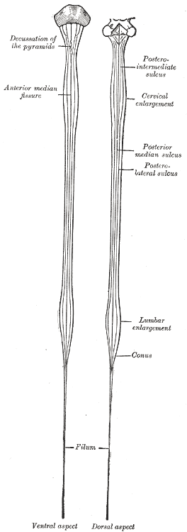 Diagrams of the medulla spinalis. (Cervical enlargement labeled at upper right.) | |
| Details | |
| Identifiers | |
| Latin | intumescentia cervicalis |
| TA98 | A14.1.02.002 |
| TA2 | 6050 |
| FMA | 74893 |
| Anatomical terminology | |
The cervical enlargement corresponds with the attachments of the large nerves which supply the upper limbs.
Located just above the brachial plexus, it extends from about the fifth cervical to the first thoracic vertebra, its maximum circumference (about 38 mm.) being on a level with the attachment of the sixth pair of cervical nerves.
The reason behind the enlargement of the cervical region is because of the increased neural input and output to the upper limbs.
An analogous region in the lower limbs occurs at the lumbar enlargement.
References
[edit]![]() This article incorporates text in the public domain from page 752 of the 20th edition of Gray's Anatomy (1918)
This article incorporates text in the public domain from page 752 of the 20th edition of Gray's Anatomy (1918)
External links
[edit]- lesson6spinalcord&coverings at The Anatomy Lesson by Wesley Norman (Georgetown University)
- Anatomy photo:02:08-0101 at the SUNY Downstate Medical Center - "Vertebral Canal and Spinal Cord: Regions of the Spinal Cord"
- Atlas image: n3a5p3 at the University of Michigan Health System - "Spinal Cord, Fetus, Posterior View"
This neuroscience article is a stub. You can help Wikipedia by expanding it. |