眼窩下孔
| 骨: 眼窩下孔 | |
|---|---|
 1 篩骨孔, 2 視神経管, 3 上眼窩裂, 4 涙嚢窩, 5 眼窩下溝, 6 下眼窩裂, 7 眼窩下孔  Articulation of nasal and lacrimal bones with maxilla. (Infraorbital foramen labeled at left.) | |
| 名称 | |
| 日本語 | 眼窩下孔 |
| 英語 | Infraorbital foramen |
| 関連構造 | |
| 上位構造 | 上顎骨 |
| 関連情報 | |
| グレイ解剖学 | 書籍中の説明(英語) |
| テンプレートを表示 | |
眼窩下孔(がんかかこう)は眼窩下管の出口。眼窩下動脈、眼窩下静脈、眼窩下神経が通る。犬歯窩上方で、眼窩下縁中央の1cm下方[1]にある。
眼窩下動脈、前上歯槽動脈、後上歯槽動脈への麻酔を目的とし、口腔内または同部の皮膚から眼窩下孔に麻酔薬を注入する眼窩下孔注射法がある[1]。
画像
-
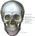 The skull from the front. (Infraorbital foramen labeled at center right, under the eye.)

脚注
- ^ a b 上山吉哉 著「第19章 麻酔 1 局所麻酔 1 局所麻酔法 5)術式 (1)上顎神経 b.眼窩下孔注射法」、白砂兼光、古郷幹彦 編『口腔外科学』(第3版)医歯薬出版、東京都文京区、2010年3月10日、741-742頁。ISBN 978-4-263-45635-4。 NCID BB01513588。
外部リンク
- cranialnerves at The Anatomy Lesson by Wesley Norman (Georgetown University) (V)
- Anatomy photo:29:os-0506 at the SUNY Downstate Medical Center (closeup)
- Anatomy figure: 22:02-08 at Human Anatomy Online, SUNY Downstate Medical Center (distance)
- Upstate.edu
- Anatomy diagram: 34256.000-1 at Roche Lexicon - illustrated navigator, Elsevier
この記事にはパブリックドメインであるグレイ解剖学第20版(1918年)158ページ本文が含まれています。
| 典拠管理データベース |
|
|---|


